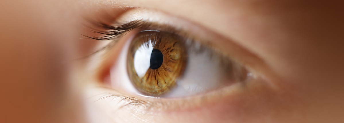
What to Expect at Your Annual Diabetes Eye Exam
Written by: Ginger Vieira
4 minute read
May 19, 2021
Your annual diabetes eye exam can take anywhere between one to two hours because there are actually four parts to the exam.
Your annual eye exam will look for signs of these five conditions, all of which can be largely prevented through managing healthy blood glucose (blood sugar) levels and maintaining an A1C at or below 7%.
- Retinopathy: Swelling and bleeding in the blood vessels of your eye’s retina that can lead to blindness
- Macular edema: Swelling and fluid build-up in the macula of your eye, often coincides with retinopathy, and can cause severe vision loss if left untreated
- Cataracts: Cloudiness in the lens of your eye that can cause vision loss
- Glaucoma: Increased fluid pressure in your eyes that if left untreated, can cause blindness
- Dry eye: Blurred vision that improves with blinking, excessive watering to compensate for the dryness and, when severe, stinging and burning in your eyes
Four types of eye exams for people with diabetes
Usually performed by an optometrist and a technician or nurse, there are four parts of a routine diabetes eye exam using different technology to look at different parts of your eye’s overall health.
Let’s take a closer look at each of these four tests.
Visual Acuity Testing
Using an eye chart and other technology, this test is likely something you’ve had done many times before if you wear glasses or contacts. It will also determine your overall vision and whether you need glasses/contacts or an update to your current prescription.
This test assesses your long-distance vision, such as when you’re driving, and your up-close vision, like when you’re reading a book.
Using different approaches, you’ll read letters and/or numbers at various distances to determine how well you can see with and without corrective lenses.
Normal vision is 20/20, which means your ability to see something from 20 feet away is consistent with people who have standard vision ability for healthy eyes. This test uses black letters on a white background and generally does not reflect your ability to see in the real world, where few things are black on white. Low contrast (greyish letters on a white background) eye charts measure vision in real world settings much more accurately and may be used by your eye specialist.
If your results come back at 20/60, for example, this means that you need to be 20 feet from something that someone with normal vision can see from 60 feet away. This helps determine the prescription for your glasses.
Diabetes can potentially lead to sudden or rapid changes in your vision and dramatic fluctuations in your eyeglass or contact lens prescription. The visual acuity test will help catch these changes.
Tonometry
This test measures the pressure in your eye, also known as intraocular pressure (IOP). Generally, normal IOP is between 10 and 21 mmHg. If your IOP measures higher than 21 mmHg, it could mean you’ve developed glaucoma (a specific form of optic nerve damage resulting in loss of vision), which can lead to blindness if left untreated. Importantly, some patients develop glaucoma even when IOP is in the normal range. Some patients never develop glaucoma despite their IOP being above 21 mmHg.
Since glaucoma generally has no symptoms until it’s severe, this test is critical to catching it early, along with a 3-D view of your optic nerves to detect early damage from eye pressure.
The test itself is painless, and simply requires you to sit still and open your eyes widely while your eye pressure is measured. While there are types of tonometers that require gentle contact with your eye using a topical anesthetic to eliminate all discomfort, some do not come in direct contact with your eye.
Dilated Eye Exam
This is the most important part of the eye exam for people with diabetes because it is the best way to catch any early signs of damage in your eyes, as well as grade the severity of any damage that has already occurred.
After applying eye drops that enlarge the pupils, giving the eye doctor a much better, 3-D view inside your eyes, you’ll probably be asked to hang out with a magazine for 15 to 30 minutes while the drops cause your pupils (the dark circle in the center of your iris) to fully open. This will let as much light into your eye as possible and prevent your pupils from getting smaller when your doctor shines a light in them. This makes it easier for your eye doctor to examine your eyes thoroughly, which is essential for detecting both macular edema and glaucoma.
Dilating your eyes means your eye doctor will be able to look into the back of your eye using non-invasive technology to detect any swelling, leakage, bleeding blood vessels, abnormal new blood vessels, nerve damage, cataracts and other eye diseases that are more common in people with diabetes.
While dilated, your vision will be blurry and you will be very sensitive to light for about two to four hours after the exam. You should bring sunglasses with you so you can get around safely and with minimal light sensitivity (photophobia) until the drops wear off. Some people may find they want a friend or family member to drive them home, especially if you already have limited vision or live far from your eye doctor’s office.
Optical Coherence Tomography
Once your eyes are dilated (as explained above), your doctor may also use noninvasive imaging technology to scan the different layers of your retina and/or the health of your optic nerve with a specialized light (sort of an optical ultrasound). This is especially important for people suspected of diabetic macular edema or glaucoma.
This test is also very useful for detecting age-related macular degeneration, a totally different but common eye disease in older adults.
To prepare for this part of your diabetes eye exam, your optometrist may put dilating eye drops in each eye. By dilating your eyes prior to the exam, it will be easier to get higher quality scans of your retina.
Your annual diabetes eye exam can feel stressful or tedious, but it is the best way to detect and prevent diabetes-related eye diseases from causing permanent vision loss or blindness.
Eye Health content is created through the ADA x BT1 Collab, with support from Focus on Diabetes™.
Related Resources

This resource on inhaled insulin was made possible with support from MannKind (makers of Afrezza (insulin...
Read more

Editor's Note: This article is about Ginger's personal experience using Afrezza, and what has worked...
Read more

Editor’s Note: We have a simple goal: tap into the power of the global diabetes...
Read more


